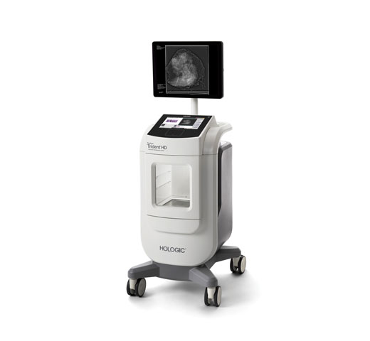
Faxitron® Core Specimen Radiography System
The system provides high resolution imaging within seconds, reducing delays for core sample verifications.1
Information for Healthcare Professionals

Overview
Documents
Immediate Core Sample Verifications
The compact unit can be located directly in the stereotactic suite, only steps from the table. With one touch of a button, a successful biopsy procedure is confirmed. Images can be instantly sent to multiple destinations without interrupting the mammography workflow.1
Automated, Accurate Image Measurements1
Automatic exposure control (AEC) and auto-calibration optimise images at the touch of a single button. They can then be sent to PACS for shared review and archiving.
Innovative CMOS Technology
The proprietary detector in the Faxitron Core system can facilitate up to 14 lp/mm resolution and achieves one of the highest image qualities in the industry.
Fully Self-Contained
The system is a self-contained unit, requiring no additional X-ray shielding.
Easy to Use
No additional training or specialised X-ray requirements are needed to operate the system.
Versatile Specimen Drawer
A built-in drawer combines with our easy to use consumable tray, allowing for transportation of the biopsy to pathology.
Unlock the Advantage of Time
The Breast Health Continuum of Care offers integrated solutions for clinical confidence, workflow efficiency and compassionate patient care. It is our purpose to give more women more time in better health.
The Faxitron Core Specimen Radiography System is part of the Hologic Specimen Radiography Solution.


1.4x Geometric Magnification2
The compact unit can be located directly in the stereotactic suite, only steps from the table. With one touch of a button, a successful biopsy procedure is confirmed. Images can be instantly sent to multiple destinations without interrupting the mammography workflow.
- Specimen imaging area: 5cm x 10cm
- Spatial resolution: 14lp/mm with fixed 1.4X geometric magnification
- External dimensions: 38cm wide x 32cm deep x 48cm tall
Insights
- Hologic Data on File. Faxitron Core V2.0 Users Manual. 04-1043-00. Rev 045, 2021
- Hologic Data on File. Cabinet X-Ray Systems MDR Technical Documentation. TFS-00054 Rev 001, 2023
Safety Data Sheets
Package Inserts
Related Products
2797
0482
Hologic BV, DA Vincilaan 5, 1930 Zaventem, Belgium
Notified Body number wherever applicable
EC Representative Information wherever applicable


